Glaucoma can injure the optic nerve while your central vision still feels normal, and the tissue lost cannot be brought back. By understanding the disease type, interpreting tests together, and matching treatment and follow-up to your risk, you can make more informed decisions about protecting the vision you have.
Key takeaways
- Lost optic-nerve tissue cannot regrow, so early diagnosis protects remaining vision.
- A complete assessment measures eye pressure, the optic nerve, visual fields, and drainage angle.
- Use glaucoma drops exactly as prescribed and attend follow-up even without symptoms.
- Severe eye pain, blurred vision, halos, nausea, and redness need urgent angle-closure assessment.
How Glaucoma Damages Vision and Which Type You May Have
Glaucoma damages the optic nerve, the cable carrying visual signals from the eye to the brain. Raised pressure, or other vulnerability at the nerve, gradually destroys nerve fibres; the resulting blind spots and visual-field loss cannot be restored because lost nerve tissue does not regrow.
Diagnostic sobriety matters: high intraocular pressure is a risk factor, not proof of disease, and normal pressure does not exclude glaucoma.
| Type | What happens | Typical pattern |
|---|---|---|
| Primary open-angle glaucoma | The drainage angle remains open but works inefficiently, allowing pressure to damage the nerve. | Usually gradual and painless |
| Angle-closure glaucoma | The iris blocks the drainage angle, causing pressure to rise rapidly or intermittently. | Often urgent; anatomy can predispose both eyes |
| Normal-tension glaucoma | Optic nerve damage occurs despite statistically normal pressure. | Field loss and nerve changes still develop |
| Secondary glaucoma | Another condition, medicine, inflammation, trauma, or eye problem raises pressure or injures the nerve. | Treating the cause matters |
Arrange earlier or more frequent assessment with increasing age, a close family history, diabetes, high myopia, steroid use, previous eye injury, or African or Asian ancestry when clinically relevant.
A sudden painful red eye with blurred vision, halos around lights, headache, nausea, vomiting, or sudden vision loss can signal acute angle-closure glaucoma. Seek emergency assessment immediately; do not wait for a routine appointment.
What a Complete Glaucoma Assessment Measures
No single intraocular pressure reading or cutoff confirms glaucoma diagnosis. High pressure is a risk factor, not proof of disease: many people with ocular hypertension never develop nerve damage, while normal-tension glaucoma can damage the optic nerve at statistically normal pressure.
A complete assessment combines several findings:
- Medical history, including diabetes, steroid use, previous eye injury, and other eye conditions.
- Family history of glaucoma and previous test results.
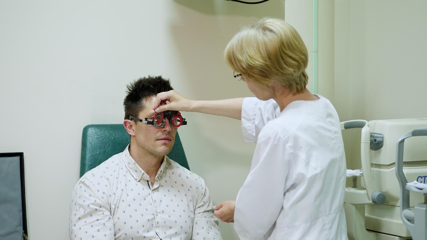
- Visual-acuity testing to measure clarity at distance and near.
- Tonometry to measure intraocular pressure.
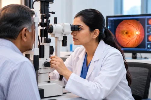
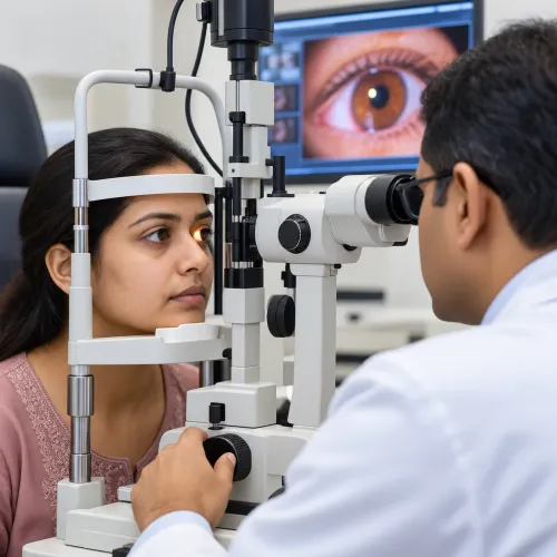
- Slit-lamp examination of the cornea, iris, lens, and anterior eye.
- Gonioscopy to inspect whether the drainage angle is open or narrowed.
- Optic-nerve evaluation for cupping, asymmetry, rim thinning, or haemorrhage.
- Retinal nerve-fibre-layer or optic-nerve optical coherence tomography (OCT) to detect structural loss.
- Standard automated perimetry to measure functional vision.
Tonometry is influenced by central corneal thickness and corneal biomechanics. A thick or stiff cornea can make the reading appear higher; a thin or more flexible cornea can make it appear lower. Interpret pressure alongside the optic-nerve appearance, OCT, and visual field.
Perimetry is a response-based test: press the button whenever you see a stimulus. False positives, fatigue, poor fixation, learning effects, and uncorrected refractive error can distort an early printout. Repeatable results that agree with examination and OCT carry more weight than one abnormal field.
When to Test, How to Prepare, and What Follow-Up Means
If you have few risk factors and normal findings, periodic assessment may be enough; glaucoma screening frequency changes when your risk or test results change. Do not choose an interval from age alone.
- Ask the clinician to set your testing schedule around family history, diabetes, high myopia, steroid exposure, suspicious optic nerves, ocular hypertension, thin corneas, or previous eye injury. These findings justify closer review than routine screening.
- Treat glaucoma follow-up intervals as individual. Existing damage, untreated pressure, visual-field and OCT change, age, life expectancy, corneal thickness, and treatment response all matter. After a new diagnosis or evidence of progression, the clinician may shorten the interval.
- Make glaucoma testing preparation easier by bringing your current glasses, every eye-drop and medicine name, prior reports and scans, steroid details, and family-history information. Tell the clinician when steroid treatment started, where it was used, and whether it continues.
- Expect a dilated eye examination when the optic nerve or retina needs a wider view. Dilating drops can temporarily blur near and distance vision and increase light sensitivity, so arrange transport if your vision remains unsuitable for driving.
At a specialist assessment in Mumbai, FOHS's K.B.H.Bachooali Charitable Eye&ENT Hospital is one local setting where your risk history, test results, and follow-up plan can be discussed together. A normal pressure reading does not cancel follow-up when the optic nerve, OCT, or visual field remains suspicious.
Related service
Glaucoma Clinic K. B. Haji Bachooali Charitable Ophthalmic and ENT Hospital, located in Parel, Mumbai, provides comprehensive glaucoma diagnosis and treatment to... View service → |
Choosing Pressure-Lowering Treatment for Open-Angle Glaucoma
Treatment for open-angle glaucoma or ocular hypertension aims to lower intraocular pressure and reduce the chance of further optic-nerve damage. It cannot replace nerve tissue already lost or restore an established visual-field defect, so the pressure target depends on your measured damage and future risk.
| Option | What it does | Main limitation or trade-off |
|---|---|---|
| Glaucoma eye drops, including prostaglandin analogues | Usually lower pressure with once-daily dosing | Redness, eyelash growth, periocular skin darkening, and potentially permanent iris darkening, especially in mixed-color irides |
| Selective laser trabeculoplasty | Improves drainage through the eye’s existing outflow pathway | The effect can fade; medicines or repeat treatment may later be needed |
| MIGS glaucoma surgery | Provides a more modest pressure reduction for selected mild-to-moderate cases | Not automatically suitable for advanced glaucoma or every cataract operation |
| Trabeculectomy or glaucoma drainage-device surgery | Provides greater pressure lowering when a low target is necessary | More intensive surgery with greater follow-up and complication considerations |
Selective laser trabeculoplasty is an established first-line option for many newly diagnosed or suspected open-angle cases, including ocular hypertension. It avoids daily dosing, but it does not guarantee permanent control.
Discuss every change in eye drops with your clinician. Prostaglandin analogues are effective, yet visible side effects can affect whether you continue treatment. A high pressure is a risk factor, not proof of glaucoma, and treatment choices must also reflect optic-nerve appearance, imaging, visual fields, age, and life expectancy.
Preventing Treatment Failure and Responding to Angle Closure
An apparently ineffective drop is often an unworkable treatment plan, not a failed medicine. Glaucoma treatment adherence matters because damage can progress while vision feels unchanged; use drops on schedule even when your sight seems normal.
1. Check the practical barriers. Missed doses, forgetfulness, cost, ocular-surface irritation, a complicated schedule, or poor eye-drop technique can reduce the amount reaching the eye. Ask the clinician to watch your technique and simplify dosing, change medicines, or address irritation.
2. Escalate pressure control when drops remain unreliable. Selective laser trabeculoplasty (SLT) can reduce pressure in many open-angle cases; its effect can fade, requiring medicines or repeat treatment. Surgery becomes relevant when pressure remains above target or adherence cannot be made workable.
3. Treat angle closure as urgent when severe pain, a red eye, blurred vision, halos, headache, nausea, vomiting, or sudden vision loss develops. Laser peripheral iridotomy creates a small opening in the iris, allowing fluid to bypass an anatomical blockage. It does not restore optic-nerve damage, and some patients still need pressure-lowering treatment and monitoring.
4. Set a personalised target intraocular pressure with your clinician. Base it on starting damage, untreated pressure, OCT or visual-field change rate, corneal thickness, age, life expectancy, family history, and other risks—not a universal “safe” number. A high pressure is not proof of glaucoma, and a statistically normal pressure is not always safe.
Frequently asked questions
How does glaucoma damage vision?
Glaucoma damages optic-nerve fibres, causing permanent blind spots and visual-field loss. Raised eye pressure is one cause, but the nerve can also be vulnerable at normal pressure.
What tests are included in a complete glaucoma assessment?
Assessment measures intraocular pressure, examines the optic nerve, tests peripheral vision, and evaluates the drainage angle. Imaging of the optic nerve and retinal nerve-fibre layer can support comparison over time.
When should you test for glaucoma?
Arrange testing when you have glaucoma symptoms or risk factors, and keep the follow-up schedule your eye specialist sets. Angle-closure symptoms require urgent assessment rather than a routine appointment.
How is open-angle glaucoma treated?
Treatment lowers eye pressure with prescribed eye drops, laser treatment, or surgery when needed. Your specialist chooses based on pressure, optic-nerve damage, visual-field results, eye anatomy, and response to treatment.
What are the warning signs of angle closure?
Severe eye pain, sudden blurred vision, halos around lights, a red eye, headache, nausea, or vomiting can signal angle closure. Seek urgent eye care instead of waiting for a scheduled visit.