Blurred vision, glare, pain, or a gritty feeling can come from a damaged or irregular cornea, but those symptoms do not identify the cause on their own. By matching symptoms and test findings to the affected corneal layer, you can understand which treatments control disease, which improve vision, and when urgent care matters.
Key takeaways
- Seek prompt care for pain, light sensitivity, redness, or a white corneal spot.
- Treatment depends on whether infection, dryness, scarring, or irregular shape causes symptoms.
- A specialist uses slit-lamp examination and targeted tests to find the cause.
- Corneal transplants are reserved for severe damage that other treatments cannot correct.
Which Corneal Symptoms Need Prompt Eye Care?
Recognise a possible corneal problem when blurred or reduced vision appears with corneal pain, light sensitivity, redness and watering, glare, halos, or a gritty foreign-body sensation. A visible white spot on the cornea is especially concerning; do not wait for a routine glasses or laser consultation.
| Warning pattern | Action | Why it matters |
|---|---|---|
| Red, painful, light-sensitive eye with a white spot or sudden vision loss | Seek emergency eye care today | Microbial keratitis can scar the cornea or cause perforation, especially in contact-lens wearers. |
| Chemical splash or penetrating injury | Rinse continuously with clean water and seek emergency care immediately | Delay allows deeper corneal damage. |
| New pain, redness, light sensitivity, or reduced vision after a corneal transplant | Contact your eye specialist urgently, even months or years later | Graft rejection can sometimes be reversed with prompt corticosteroid treatment; delay can cause graft failure. |
Arrange prompt assessment for changing glasses prescriptions, night-vision difficulty, glare, or ghost images, which can signal keratoconus. Avoid rubbing your eyes and control allergy-related itching. Morning blur can occur with Fuchs’ dystrophy; persistent blur or painful fluid-filled bullae require specialist review. Do not use leftover antibiotic or steroid drops.
Which Corneal Conditions Require Different Treatments?
Keratoconus, infection, dystrophy, scarring, dry-eye damage, and injury need different treatments because each affects a different part of the cornea: its shape, tissue, surface, or clarity.
| Condition | Treatment focus | Why it differs |
|---|---|---|
| Keratoconus | Glasses or specialty lenses; corneal collagen cross-linking for documented progression | Cross-linking strengthens the cornea and slows change, but does not reliably remove existing irregular astigmatism. |
| Infection: keratitis or corneal ulcer | Urgent examination, possible culture, and frequent prescription antibiotic, antifungal, or antiviral treatment | Delay can leave a scar or cause perforation, especially when a contact-lens wearer has pain, redness, light sensitivity, or a white spot. |
| Corneal dystrophy | Surface measures for early disease; PTK for selected superficial disease; endothelial keratoplasty for advanced Fuchs’ dystrophy | Fuchs’ swelling can cause morning blur, while painful bullae or persistent loss may require replacement of the damaged endothelial layer. |
| Corneal scarring | PTK for selected superficial scars; deeper scars may need another procedure or a transplant | PTK cannot safely remove every opacity and can cause haze or refractive change. |
| Dry-eye damage | Lubricating drops and treatment of the tear-film problem | Restoring moisture allows surface cells to heal. |
| Injury | Immediate irrigation for chemical exposure, removal of foreign bodies, or repair of deeper wounds | Treatment depends on the cause and depth; a penetrating injury is an emergency. |
LASIK is unsafe for established keratoconus, abnormal thickness, or significant dry eye. Topography, tomography, and pachymetry guide that decision.
How Does a Specialist Identify the Cause of Blurred Vision?
A glasses test shows whether blur is refractive, but it cannot explain every cause. A specialist compares your best-corrected vision with the cornea’s shape, thickness and surface, then checks the lens, retina and optic nerve so treatment targets the real problem.
| Examination | What it shows | Clue it provides |
|---|---|---|
| Refraction | The prescription needed for clearest vision | Blur from myopia, astigmatism or another focusing error |
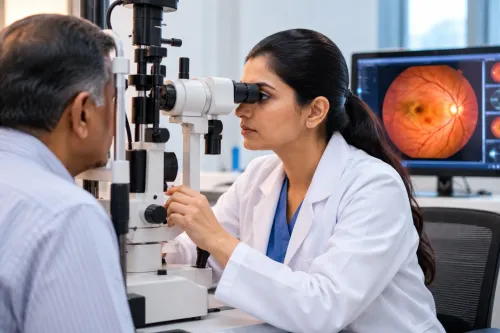
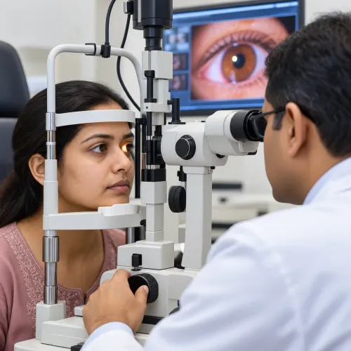
| Slit-lamp examination | The cornea under magnification, plus the lens and front of the eye | Scratches, scarring, swelling, dry-eye damage, cataract or visible inflammation |
| Corneal topography | A map of the front corneal surface | Irregular astigmatism, keratoconus or an uneven scar |
| Corneal tomography | Front and back corneal surfaces with a three-dimensional thickness profile | Structural weakness or ectasia that a front-surface map might miss |
| Pachymetry | Corneal thickness at measured points | Abnormally thin or swollen tissue, and safety information before corneal procedures |
Tear-film assessment looks for unstable tears, blepharitis and surface disease that cause fluctuating blur or contact-lens intolerance. Burning does not prove infection, so a clinician may stain the cornea with fluorescein and collect a sample for laboratory testing when an ulcer, keratitis or unusual discharge is present.
At FOHS's K.B.H.Bachooali Charitable Eye&ENT Hospital, asking whether the examination included retinal and lens checks is useful when corneal scans do not explain the reduced vision. A normal corneal map does not rule out cataract, retinal disease or glaucoma.
Related service
Cornea Clinic K. B. Haji Bachooali Charitable Ophthalmic and ENT Hospital, located in Parel, Mumbai, offers comprehensive diagnosis and treatment for a wide range... View service → |
Which Medicines and Surface Treatments Can Help?
Surface treatment can restore clearer, more comfortable vision when dryness, inflammation, infection, or a superficial defect is causing blur. The right medicine depends on the cause; using steroid or leftover antibiotic drops without an examination can worsen an infection.
| Option | Appropriate when | What it improves |
|---|---|---|
| Lubricating eye drops | Dry eye, blepharitis, allergy, or tear film instability causes burning, fluctuating blur, or lens intolerance | Moisture, comfort, surface smoothness, and visual stability |
| Antibiotic eye drops | A clinician suspects bacterial keratitis or a corneal ulcer | Bacterial infection and the risk of deeper damage |
| Antifungal treatment | Examination and testing point to fungal keratitis, often after plant material injury | Fungal growth and progressive corneal destruction |
| Antiviral treatment | Herpes simplex or another viral corneal infection affects the surface | Viral activity, pain, and recurrent epithelial breakdown |
| Bandage contact lens | A recurrent erosion or superficial epithelial defect needs protection after infection is excluded or treated | Healing comfort by shielding exposed nerve endings |
A red, painful, light sensitive eye with a white corneal spot needs same day ophthalmic assessment, especially in a contact lens wearer. Doctors may prescribe frequent antimicrobial drops and take a culture. A bandage lens requires professional fitting and review because it can conceal or worsen infection.
How Are Irregular Corneas and Reduced Vision Corrected?
Glasses are the better first route when refraction corrects the blur and the corneal shape is regular. If glasses leave ghosting or distorted vision, a specialist may fit a lens that creates a smoother optical surface.
| Option | What it improves | When it is the better route |
|---|---|---|
| Glasses | Regular short-sightedness, long-sightedness, or astigmatism | The cornea is stable and spectacles provide clear, comfortable vision |
| Rigid gas-permeable lenses | Irregular astigmatism by creating a regular front surface | Keratoconus or corneal irregularity causes blur that glasses cannot correct |
| Scleral lenses | Vision and comfort by vaulting the cornea over a fluid reservoir | A highly irregular or sensitive cornea needs stable optics; fitting, cleaning, and monitoring are essential |
| Corneal cross-linking | Corneal strength, not immediate optical clarity | Documented keratoconus progression requires slowing or stopping further bulging; glasses or lenses may still be needed |
| Phototherapeutic keratectomy | Selected superficial scars, recurrent erosions, or anterior dystrophy deposits | The opacity or roughness is superficial and treatable; haze, refractive change, deep scarring, or thinning can make PTK unsuitable |
Laser procedures such as LASIK or PRK suit carefully selected eyes with a stable, regular cornea and adequate thickness. They are a poor choice for progressive keratoconus, significant thinning, or irregular scarring because removing tissue can worsen structural weakness.
Do not treat corneal blur by assumption. Refraction, slit-lamp examination, and corneal mapping must confirm that the cornea—not the lens, retina, glaucoma, or dry eye—is the source.
When Is a Corneal Transplant the Right Option?
A corneal transplant is appropriate when damaged tissue prevents useful vision or causes pain that other treatments cannot control. The surgeon selects the graft according to the affected corneal layer.
| Technique | Tissue replaced | Recovery and trade-off |
|---|---|---|
| DALK | Front and middle layers, preserving the healthy endothelium | Avoids endothelial rejection, but vision can take time to settle |
| DMEK | A very thin layer of diseased posterior endothelium | Usually faster recovery and fewer suture problems; a detached graft may need a rebubble |
| DSAEK | Posterior endothelial tissue with a supporting layer | Faster recovery than full-thickness surgery, with possible rebubble treatment |
| Penetrating keratoplasty | The full corneal thickness | Recovery can take many months, with astigmatism often requiring glasses or contact lenses |
Expect prescription corticosteroid drops and other medicines exactly as directed. Do not stop steroid drops without medical advice; treatment plans can last months or longer.
- Attend frequent early examinations, then long-term reviews, because graft failure can follow infection, glaucoma, endothelial cell loss, or rejection.
- Treat new redness, light sensitivity, pain, or reduced vision as urgent warning signs. Prompt examination and corticosteroid treatment can sometimes reverse rejection.
- Understand that a graft does not remove cataract, retinal disease, or every refractive error; further treatment or corrective lenses may still be necessary.
Frequently asked questions
Which corneal symptoms need prompt eye care?
Seek prompt assessment for blurred or reduced vision with pain, light sensitivity, redness, watering, glare, halos, or a gritty sensation. A white spot on the cornea needs urgent attention.
How does a specialist identify the cause of blurred vision?
An eye specialist examines the cornea with a slit lamp and may use corneal mapping, tear-film tests, fluorescein staining, or infection testing.
Which medicines and surface treatments can help corneal problems?
Treatment depends on the cause and can include lubricating drops, antibiotic or antiviral medicines, anti-inflammatory treatment, protective lenses, or treatment for eyelid and tear-film problems.
When is a corneal transplant the right option?
A transplant is considered when corneal scarring, swelling, thinning, or structural damage causes significant vision loss that cannot be corrected with medicines, lenses, or other procedures.